
Video dermatoscopy
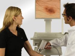
Video dermatoscopy is a diagnostic method based on the same principle as dermatoscopy. The basic difference is that the dermatoscopic digital images are taken, stored and compared with images from the patient’s next visit on a computer screen. Due to computer software, it is possible to archive images and follow individual history of each patient. Computer-assisted analysis of individual nevi enables magnified observation of the lesion, archiving the images and a follow-up examination with comparison. Video dermatoscope (digital dermatoscope) is a connection of a dermatoscope, computer software and a digital camera.
Before dermatoscopic examination, a patient should inform the doctor about duration of the skin nevi, the rate of its growth, its initial colour (red, brown, etc.), spontaneous painfulness of the nevi or pain upon compression.
Depending on the diagnosis (based on the patient's history, location of the nevi, analysis and results of dermatoscopic examination) the doctor suggests removal of particular pigmented lesions or their observation until a follow-up consultation.
FotoFinder Dermoscope 800 HD by FotoFinder Systems GmbH
The device allows for the performance of non-invasive and pain-free examinations of patients. During the diagnosis, it is possible to precisely evaluate the risk of skin cancer in a certain patient as well as initiate monitoring of the patients belonging to the group of patients that are at higher risk of developing a skin cancer: numerous acquired nevi, classic atypical mole syndrome, FAM-M syndrome and positive personal or family history of melanoma. It enables a doctor to undertake the right decision about the necessity and course of treatment.FotoFinder
Unique features of the FotoFinder Dermoscope 800 HD.
1. Long-term observation and archiving - due to the new 800 HD camera and proprietary lens.
2. Immediate screening of numerous lesions in microscope mode, without the need of creating individual patient's database and record.
3. Designed especially to meet the needs of dermatoscopy laboratories, a powerful patient database, saves data and images directly in the patient's chart, allows for instant access, identification and parallel analysis of baseline and all indicated follow-up images.
4. Assessment of microcirculation - capillaroscopy test.
DIAGNOSTIC PROGRAMS
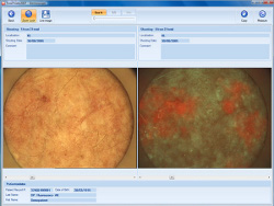
Moleanalyzer performs computer-assisted analysis of melanocytic nevi, evaluation of eventual atypia and differentiating with benign lesions found on the skin. The analysis of melanocytic nevi is made according to clinically approved pattern recognition algorithms. The obtained malignancy score helps the doctor to make diagnosis and decisions concerning further treatment. Moleanalyzer has been developed in cooperation with the Department of Dermatology at the German University of Tübingen, under the supervision of Prof. Dr. med. Claus Garbe.
The examination step by step:
1. The camera of video dermatoscope is placed over the nevi on the skin.
2. The image of the nevi is displayed on computer screen, magnified 20-70 times.
3. The software performs detailed, algebraic analysis of the parameters within the nevi (e.g. pigment distribution, edges, shape, etc.).
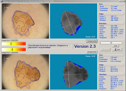
4. The obtained malignancy score helps the doctor to make decision about excision of the nevi.
5. The patient's data are archived in the computer memory.
Basic advantages of Moleanalyzer include:
- possibility of detection of minimal changes in the size, pigmentation, structure and borders of the nevi,
- early and precise diagnosis of malignant melanoma,
- high levels of sensitivity - 92%,
- second-opinion for doctors, helpful in making decision about the way of treatment: surgical excision, laser therapy, photodynamic therapy, etc.,
- creation of a digital image database, containing the analysed images of skin nevi,
- the examination is painless, non-invasive and does not require earlier preparation.
Bodyscan pro - provides computer archiving and enables observation of patients with multiple melanocytic nevi (computer comparative analysis). Thanks to computer-aided diagnosis, the risk of overlooking of new lesions on the skin can be reduced to a minimum. The expert system automatically detects new lesions and compares the current body photos of the same area with those from the last examination, which is especially important for the patients with atypical mole syndrome.
The examination step by step:
1. A patient stands on a special mat, at a determined distance from the digital camera.
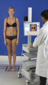
2. A series of photographs is taken in various body postures.
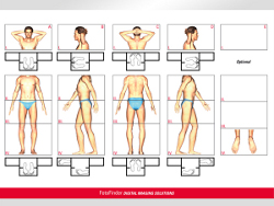
3. The system Bodyscan compares the current nevi photos with those from the past examinations (e.g. 6 months earlier).
4. Comparative analysis shows the recently developed moles on the skin...
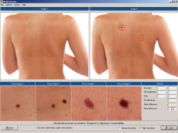
5. ...as well as it detects structural changes within the individual nevi already existing during the previous examination.
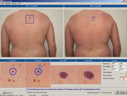
6. The obtained results allow the doctor to make a decision whether particular moles should be excised or left for further observation.
Basic advantages of Bodyscan pro include:
- comparison of macro- and dermatoscopic images of all skin lesions mapped over time (e.g. after a follow-up consultation 6 months ago),
- observation of melanocytic nevi evolution,
- detection of new moles occurring on the skin over time.
Fluorescence diagnostics (FD) serves qualitative and quantitative photodynamic diagnosis (PDD) of basal-cell carcinomas, squamous cell carcinomas (SCC) and benign skin cancers.
The examination step by step:
1. A photosensitizer is applied on the skin lesion - it accumulates in the tumour cells. When viewed through the special lens from FotoFinder, the photosensitizer fluoresces red and the tumour can be differentiated from the healthy skin.
2. The camera of video dermatoscope with a special attachment - FD lens is placed over the skin.
3. Computer analysis of the lesion is performed.
Primarily, the innovation of FD lens is a precise determination of the tumour boundaries, which prevents recurrence and spreading of cancer. The method is particularly useful in cases of large tumours, but also in cases of small tumours localised around eyelids, nerves or arteries. No other diagnostic method may assure such precision in determining the boundaries of tumour tissue.
Basic advantages of FD lens:
- enables exact determination of a lesion's location and its boundaries, so that it may be precisely removed with a minimal margin of healthy tissue,
- indicates present transit metastasis or satellites,
- serves as a tool for monitoring the effectiveness of any therapeutic method (e.g. chemotherapy, radiotherapy, PDT or surgical intervention),
- assures possibility of many examinations without side-effects (unlike other diagnostic methods),
- enables removal of a particular lesion with a photo dynamic therapy (PDT);
Trichoscan - a tool for digital qualitative and quantitative hair analysis, used for diagnostics in hair loss.