
FotoFinder
Video dermatoscope
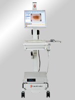 Video dermatoscope FotoFinder Dermoscope 800 HD, produced by a German FotoFinder Systems GmbH, is a computerised system of pigmented nevi (moles) mapping, which allows doctors to prepare a complete image documentation of individual patient's moles. The device allows for the performance of non-invasive and pain-free examinations of patients. During the diagnosis, it is possible to precisely evaluate the risk of skin cancer in a certain patient as well as initiate monitoring of the group of patients that are
at higher risk of developing a skin cancer: numerous acquired nevi, classic atypical mole syndrome, FAM-M syndrome and positive personal or family history of melanoma. It allows for immediate implementation of a proper treatment.
Video dermatoscope FotoFinder Dermoscope 800 HD, produced by a German FotoFinder Systems GmbH, is a computerised system of pigmented nevi (moles) mapping, which allows doctors to prepare a complete image documentation of individual patient's moles. The device allows for the performance of non-invasive and pain-free examinations of patients. During the diagnosis, it is possible to precisely evaluate the risk of skin cancer in a certain patient as well as initiate monitoring of the group of patients that are
at higher risk of developing a skin cancer: numerous acquired nevi, classic atypical mole syndrome, FAM-M syndrome and positive personal or family history of melanoma. It allows for immediate implementation of a proper treatment.
Unique features of the FotoFinder Dermoscope 800 HD:
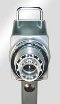 1. Long-term observation and archiving. - due to the new 800 HD camera and proprietary lens.
2. Immediate screening of numerous lesions in microscope mode, without the need of creating individual patient's database and record.
3. Designed especially to meet the needs of dermatoscopy laboratories, a powerful patient database, saves data and images directly in the patient's chart, allows for instant access, identification and analysis of baseline and all indicated follow-up images.
Specialistic software interacting with the Fotifinder Dermoscope:
Moleanalyzer - for the early detection of malignant melanoma. It allows for the observation of melanocytic nevi, comparing images taken within a given period of time. In addition, the software offers a parallel analysis of two moles side-by-side, comparison of parameters and classification of lesions into benign and malignant, according to clinically approved pattern recognition algorithms.
 Bodyscan pro - for the analysis of numerous moles. Bodyscan pro software automatically compares baseline and follow-up images of the whole body on-screen. The new or changed lesions are marked with a red circle. It's a precise, time-saving technique, intended especially for patients with dysplastic nevus syndrome.
Trichoscan - for hair structure analysis. Trichoscan is a method combining epiluminescence microscopy with digital image analysis. It allows, among others, for determining the anagen-telogen-ratio of hair growth. Trichoscan is a modified version of the traditional trichogram. The application works automatically, is completely painless and determines the main parameters of hair growth. It helps to diagnose certain types of balding and evaluate the results of different hair therapies.
 Fluorescence diagnosis (FD) is a proven method of detecting non-melanoma skin cancer and cutaneous precancerous stages such as solar keratoses and basal cell carcinomas. It can be combined with photodynamic therapy (PTD) or followed by a surgical intervention. The FD lens is easily integrated with a camera, while the treatment room may be darkened or the Wood's-light can be applied.